

Overview of HIF (Humeral Intercondylar Fissure)
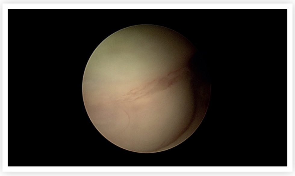
HIF is a fissure (fine crack) that separates the two sides of the humeral condyle. The fissure is believed to develop as a ‘stress-fracture’ caused by repeated uneven loading of the humeral condyle. This leads to microscopic and latterly macroscopic separation from the articular surface up towards the humeral fossa. Indeed, Danielski et al (2022) revealed a cartilage lesion on the caudal aspect of the humeral condyle in dogs with these lesions, suggesting uneven loading of the joint and hinting at a cause.
There is a physis present at this location in the growing youngster, but this tends to fuse at around four to five months of age. In the past it was thought that this physis never fused and this was termed Incomplete Ossification of the Humeral Condylar (IOHC). Observations by Moores et al (2017) revealed that many of these lesions develop over time from a previously normal, fused, condyle. Hence the currently used term HIF in adult patients.
HIF can be unilateral or bilateral and their presence predisposes the area to spontaneous fracture. HIF is seen most in commonly in Spaniels breeds, particularly English Springer Spaniels.

Signs
Signs can be vary from mild intermittent lameness, to marked persistent forelimb lameness and dogs tend to be painful when the elbow is extended during examination.
These signs warrant consideration for further tests, particularly in predisposed breeds.
Diagnosis
Examination of the patient will reveal elbow pain on manipulation and often muscle wastage on the affected leg. Conventional radiography can be used to diagnose a HIF lesion although this is not an overly sensitive diagnostic tool since the fissure can be oblique to the radiograph plane, and so can be missed. Computed tomography (CT) is considered the gold standard for diagnosis and offers the opportunity of creating a 3D guide for intercondylar screw placement.

Treatment
The goal of treatment is to stabilise the bone either side of the fissure to prevent pain from abnormal movement but also potential for fracture. A transcondylar screw is placed across the condyles preventing separation/fracture of the humeral condyles. This may be placed in ‘lag’ fashion which pulls the condyles together. Occasionally a lateral plate may be placed to further protect the screw from forces placed through the elbow joint.Complications include seroma, implant infection and implant failure. Appropriate post operative care will help minimise these risks.
Most dogs who have HIF lesions treated with transcondylar screws return to normal pain free function. Some may also be suffering from concurrent elbow disease, such as medial coronoid (see Elbow Dysplasia info).
- Danielski A, Yeadon R. Humero-anconeal elbow incongruity in spaniel breed dogs with humeral intracondylar fissure: Arthroscopic findings. Vet Surg. 2022 Jan;51(1):117-124. doi: 10.1111/vsu.13728. Epub 2021 Sep 28. PMID: 34581450; PMCID: PMC9293410.
- Moores AP, Moores AL. The natural history of humeral intracondylar fissure: an observational study of 30 dogs. J Small Anim Pract. 2017 Jun;58(6):337-341. doi: 10.1111/jsap.12670. Epub 2017 Mar 29. PMID: 28369948.
