

When is surgical treatment for a corneal ulcer indicated?
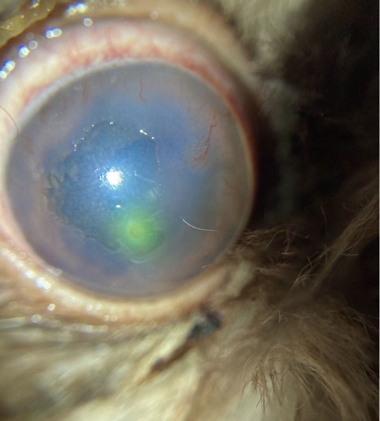
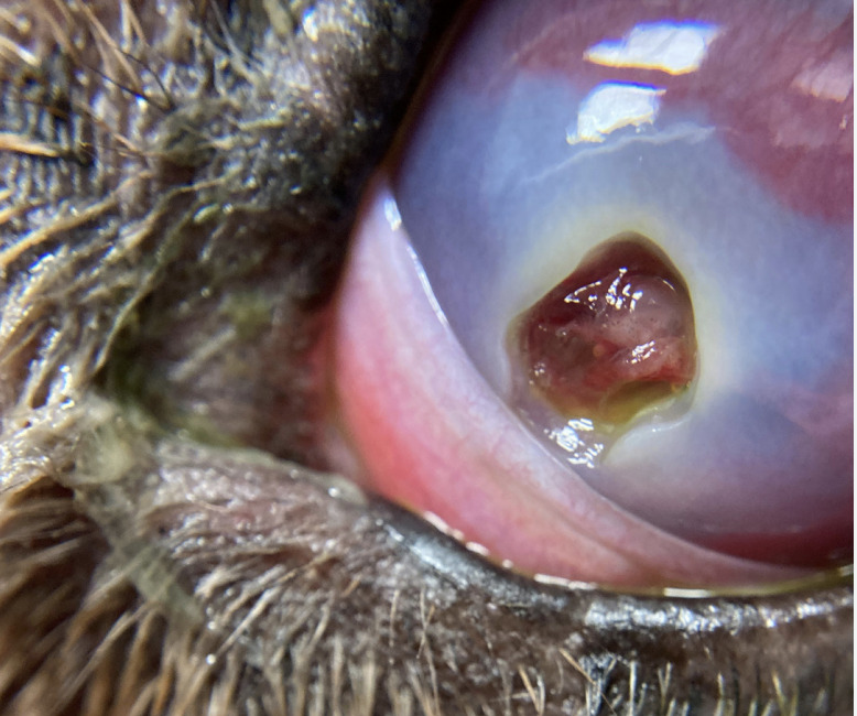
Shallow corneal ulcers typically do not require surgical intervention and may be managed medically, whereas medium to deep lesions—particularly those exhibiting melting keratitis—are strong indications for surgery. In patients with predisposing factors, such as an overly large palpebral fissure, early surgical management is recommended. Procedures may be supplemented with a medial canthoplasty to reduce exposure and improve healing.
Surgical treatment of corneal ulcers serves several critical purposes: it allows for debridement of necrotic tissue, provides structural support to the compromised cornea, and facilitates vascularization, bringing blood supply closer to the affected area to promote healing and reduce the risk of complications.
Surgical management
The surgical treatment of choice for corneal ulcers is typically either a conjunctival graft or a corneal graft. Several types of conjunctival grafts are available, including pedicle, bridge, island, and hood grafts. The type of graft used is selected based on the size, location, and nature of the lesion.
Structurally, a corneo-conjunctival graft offers superior support compared to conjunctival grafts alone, as it advances vascular supply directly toward the ulcerated area, promoting more effective healing. Whenever possible, incorporating the vascular response into the graft enhances the reparative process.
An additional advantage of corneal grafting is that it can provide a clear repair, which is particularly important when the lesion involves the visual axis, helping to preserve vision while restoring corneal integrity.
Post operatively patients will require a plastic elizabethan collar to prevent self trauma and damage to the surgical site. Occular medications such as lubricants, antibiotics and oral analgesia may also be prescribed.
Good quality ophthalmic instruments, magnification and fine suture material are essential for conjunctival grafts.
Corneal grafts require fixed depth knives and lamellar blades to harvest a graft as well as well as magnification and fine suturing technique to position the graft.
You can read more about treatment and aftercare in our resource section here.
