
Presentation
Patient Information:
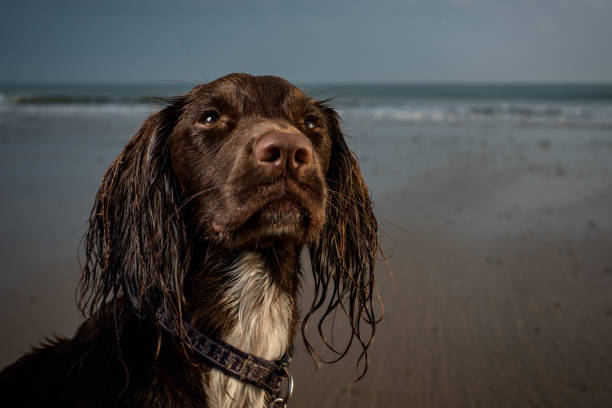
Name: Sam
Age: 6 years 5 months
Breed: Springer X Cocker Spaniel
Weight: 25.9 kg
Sex: Entire Male
Body Condition Score (BCS): 5/9Clinical Presentation and diagnostics:
Sam presented as an urgent referral with a one-hour history of acute left forelimb lameness graded at 10/10. The referring practice, under general anaesthesia, observed elbow pain and obtained radiographs which demonstrated fractures to both
humeral condyles.
Planning and Surgical Intervention:
Bi-condylar fractures, such as the one encountered in Sam’s left elbow, necessitate meticulous anatomical reconstruction to ensure optimal joint function.
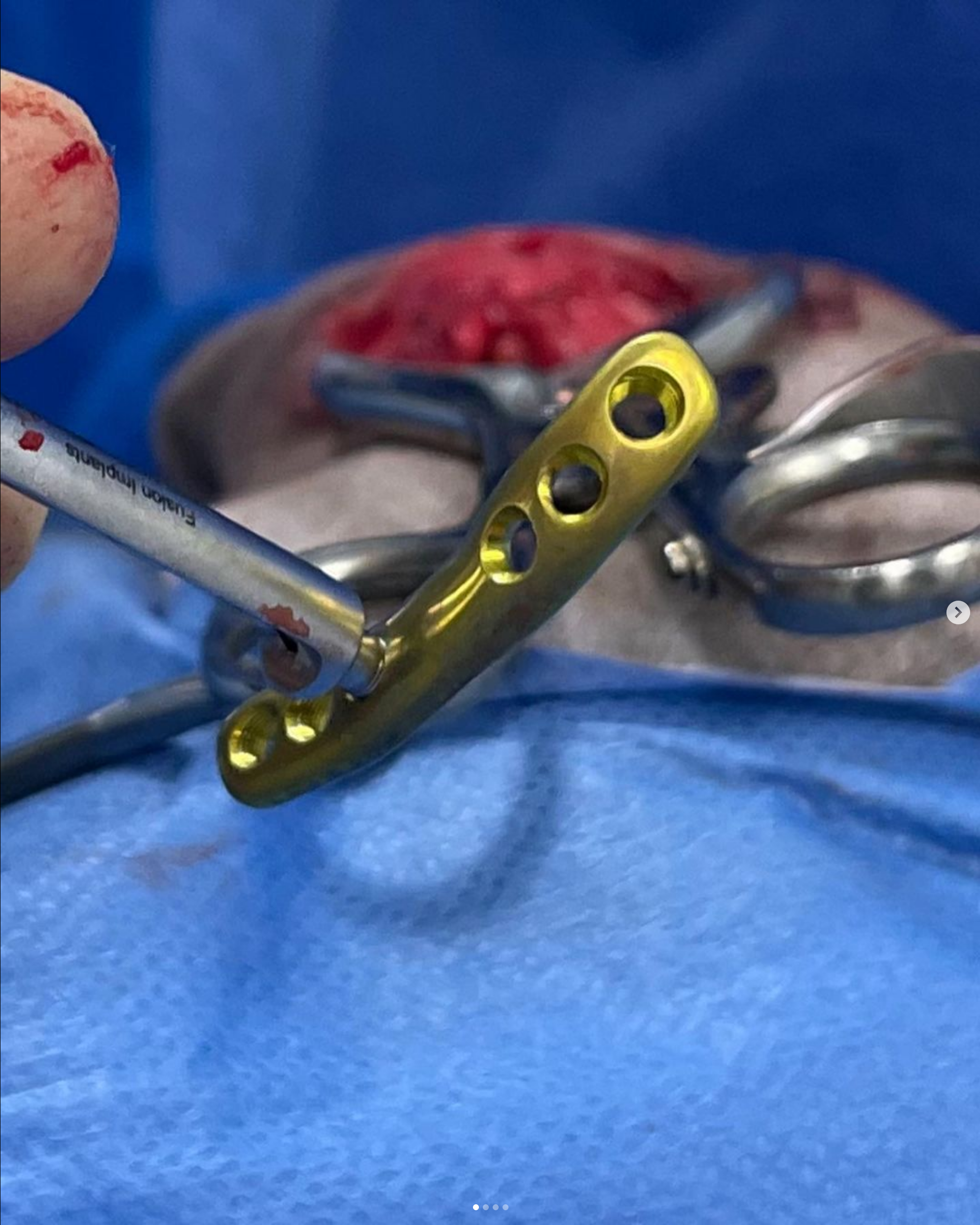
Addressing these fractures requires two separate surgical approaches (lateral and medial). The chosen surgical method of fixation for Sam involved bilateral plating and transcortical screw fixation.
Anticipated complications encompassed infection, potential nerve trauma (with irreversible implications), non-union, implant failure and osteoarthritis.Surgery:
Two of our Rata Surgeons (Matt and Joe) attended for Sam’s surgery 24 hours after his fracture – early intervention in articular fractures aligns with the AO principle of early return to function in joint fractures. This directly results in better outcomes.Sam was premedicated with medetomidine (10ug/ug) combined with methadone (0.3mg/kg), induced with propofol via a saphenous IV catheter and maintained on Isoflurane. Intravenous Paracetamol was administered at 20mg/kg q 8hrs and regional anaesthesia was applied with bupivicaine 0.5% at the Radial (lateral), ulnar, median, and musculocutaneous nerves (medial) aka a RUMM block.
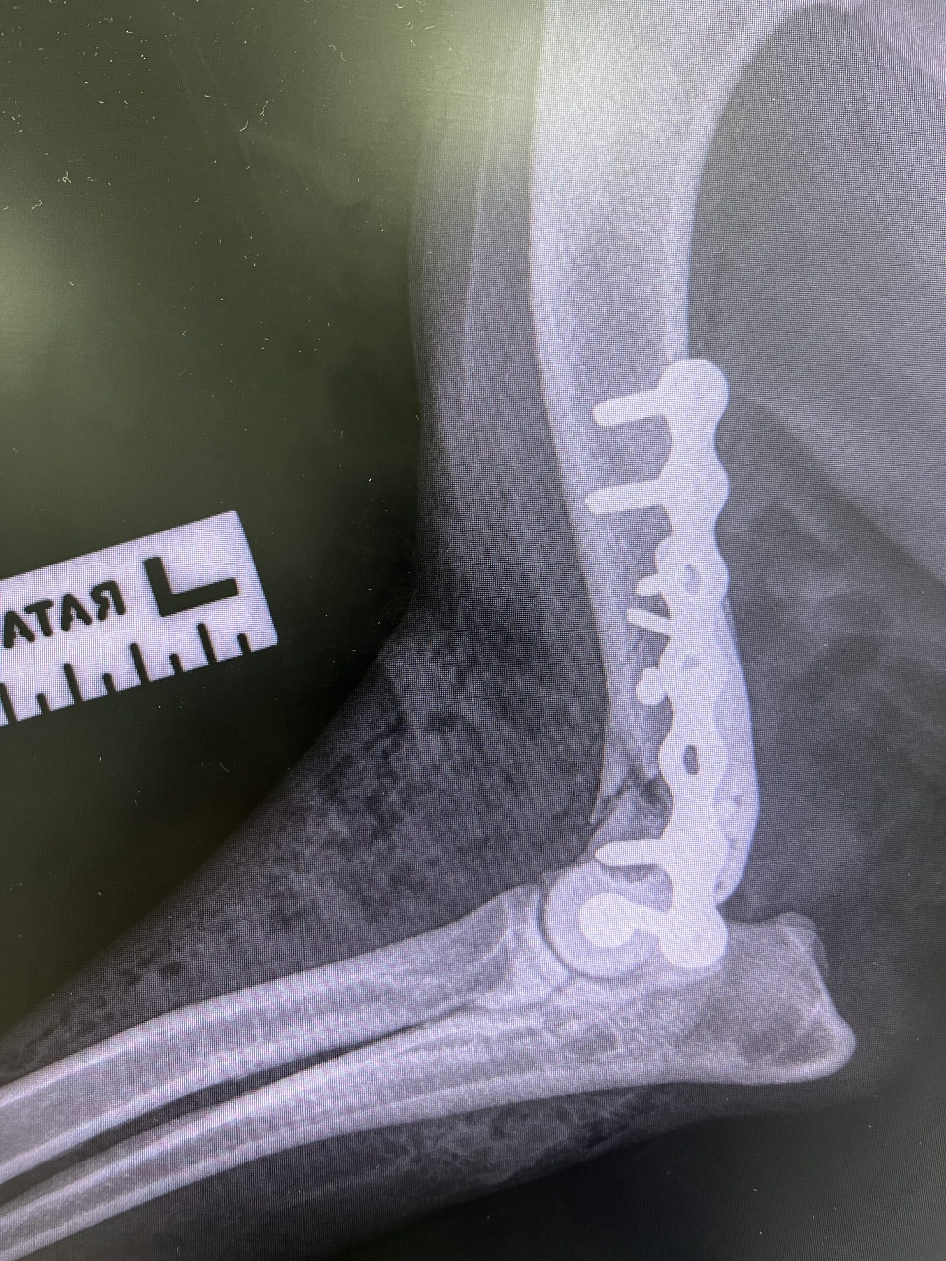
 Cranial Caudal immediate post operative radiograph
Surgery
Medial approach:
The medial approach is aimed at reconstructing the medial condyle – the first step of the
procedure. It commenced with a skin incision centered on the medial humeral epicondyle.
Subsequent dissection through subcutaneous fat and fascia (preserving neurovascular
structures) led to the exposure of the deep antebrachial fascia. Preservation of the ulnar nerve, collateral ulnar artery and vein, median nerve and brachial artery and vein was imperative.The comminution present was addressed by holding the fragment in reduction with point – to – point forceps and stablising with two 2.7 mm cortical screws placed in lag fashion. A temporary pin was placed to maintain reduction of the medial condyle, followed by stabilization with 3.5 Evolox(N2) plate (offering robust fixation and flexibility of locking screw placement).Lateral Approach:
The lateral approach was aimed at reconstructing the lateral epicondyle – the second step of the procedure. The landmarks for the incision were identified through palpation of the lateral head of the triceps muscle, lateral epicondyle, and olecranon. The incision extended along the humerus exposing the deep brachial and antebrachial fascia.
Identification and preservation of the radial nerve is a key step to this approach. The centre of the humeral condyle was rotated on the medial collateral ligament to allow visualisation of the fracture surface. This was debrided of fibrous tissue before incrementally larger drill holes were made from inside to outside. The condyles were reduced, a centered drill guide applied to the 4.5mm pilot hole and a 3.2mm pilot hole was drilled into the medial condyle. The hole was measured for depth, the start of the trans cortex tapped and a self – tapping 4.5mm screw placed and tightened in lag fashion. The repair was augmented with a lateral plate – for this a Fusion LEAP plate was used.The surgical techniques employed in Sam’s case align with AO principles; emphasizing
anatomical reduction, stable fixation, and early mobilisation. The use of lag screws, temporary k-wires, and anatomical plates adheres to the AO concept of achieving fracture stability through biomechanically sound constructs. The choice of implants, such as the Evolox(N2) plate and Fusion LEAP, reflects the AO principle of selecting implants that offer stability while minimising invasiveness.
Post operative care
Post-Operative Care:
Following the surgical intervention, Sam underwent a six-week period of strict cage rest. Initial
reports were encouraging, with Sam displaying comfort and apparent lameness-free walking.
However, the identification of a visible cranial fragment raised concerns, prompting a careful
approach to the subsequent phases of Sam’s recovery.
Advice to Owners:
During the six-week post-operative period, Sam’s owners played a crucial role in ensuring his
well-being. Strict adherence to cage rest was paramount to facilitate the initial stages of
healing. While Sam’s comfort was evident, the presence of the cranial fragment necessitated a
cautious approach.
Six-Week Follow-Up:
At the six-week mark, radiographs provided valuable insights into Sam’s progress. The images
revealed stable implants with noticeable callus calcification around the distal humerus and
fracture sites. This positive development validated the effectiveness of the surgical intervention
and indicated the initiation of the healing process.
Reassessment and Gradual Transition:
With the six-week follow-up, Sam’s overall condition was reassessed. Reports indicated his
well-being, and the gradual transition from confinement to increased leash walking was advised
over the next six weeks. This transitional phase aimed to strike a balance between promoting
controlled mobility and ensuring continued healing.
Positive Signs of Healing:
The radiographic union observed at the six-week mark provided tangible evidence of Sam’s
healing journey. The initially identified cranial fragment, while a concern in the early stages,
exhibited signs of integration and alignment with surrounding tissues. This positive development
underscored the body’s natural ability to respond to the surgical intervention and hinted at a
favorable prognosis.
Future Expectations
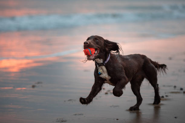
Looking ahead, Sam’s recovery trajectory is optimistic. Owners were informed that the
complete healing process might extend up to 12 weeks, and ongoing monitoring would be
essential. The collaborative effort between the veterinary team and the owners remains
instrumental in ensuring Sam’s continued progress and a successful return to normal function.Continued Follow-Up:
As Sam’s recovery advances, continued follow-up appointments will be scheduled to monitor
his overall well-being and assess the evolution of the healing process. The commitment to
comprehensive care extends beyond the immediate post-operative period, emphasizing the
importance of ongoing collaboration between veterinary professionals and dedicated pet
owners.
Next Steps:
Stay tuned for upcoming videos and further updates documenting Sam’s recovery journey. Our
commitment to comprehensive care ensures ongoing monitoring and adjustments to his
rehabilitation plan as needed
