

Optimising outcomes after tumour removal in pets
Following a tumour diagnosis, our primary goal is always to achieve the best possible outcome for our canine or feline companions, often through surgical removal of the neoplasm. However, in some cases—due to the size or location of the tumour—excision may leave a significant defect that cannot be closed primarily (where the skin edges are brought together directly). This is where advanced skin reconstruction techniques prove particularly useful, enabling not only functional repair of the defect but also a superior cosmetic result compared to open wound healing.
Why is skin reconstruction so important?
- Optimal Tumour Excision: Tumour removal often requires wide surgical margins to ensure a “clean” excision (free of cancerous cells). This can result in significant skin defects that cannot be simply closed directly. Reconstruction enables us to prioritise complete tumour removal without compromising our ability to close the wound.
- Preventing Complications: Large open wounds can take a long time to heal and are prone to infection, and bandaging issues. Successful reconstruction minimises these risks by providing a tension-free, healthy tissue closure.
- Restoring Function and Mobility: Depending on the tumour location, second-intention healing can lead to scar tissue formation that may severely limit a pet’s movement or comfort. Reconstruction allows us to restore normal function and prevent chronic pain or irritation.
- Enhancing Quality of Life: A well-reconstructed skin defect will have a better cosmetic appearance compared to a wound that heals by second intention, as well as healing more quickly, enabling our patient to return to normal activities sooner and significantly improving their quality of life after surgery.
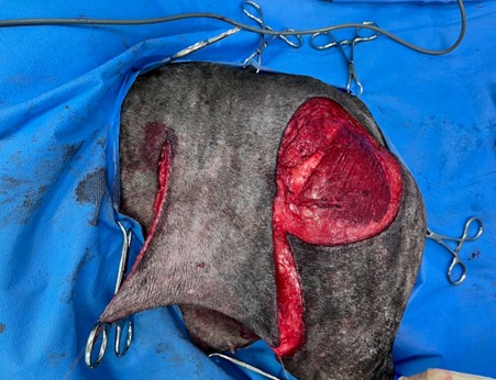
Advanced skin reconstruction techniques
-
-
Local Flaps: These involve mobilising adjacent skin and subcutaneous tissue before rotating or advancing it to cover the defect. Examples include:
-
Advancement Flaps: Moving a rectangular or V-shaped flap directly into the defect.
-
Transposition/Rotation Flaps: Pivoting an adjacent flap of skin into the defect.
-
Interpolation Flaps: Moving a rectangular flap from a non-adjacent site over an intact skin bridge to the defect.
-
Axial Pattern Flaps: These flaps maintain a vascular connection to their original site while the rest of the flap is transferred to a distant defect. Common examples include the caudal superficial epigastric flap and the thoracodorsal flap. These flaps of skin have their own direct blood supply, making them very robust.
-
Skin Grafts: These involve harvesting a thin layer of skin (split-thickness or full-thickness) from a donor site and transplanting it to the defect. Grafts rely on the recipient bed for vascularisation and are very delicate by nature, so the recipient site has to be carefully immobilised and bandaged for several weeks whilst the graft heals.
-
Tissue Expanders/Skin Stretching Devices: Used more rarely, these can be surgically implanted or glued to the skin to facilitate gradual stretching of skin adjacent to the tumour, creating additional tissue for reconstruction.
-
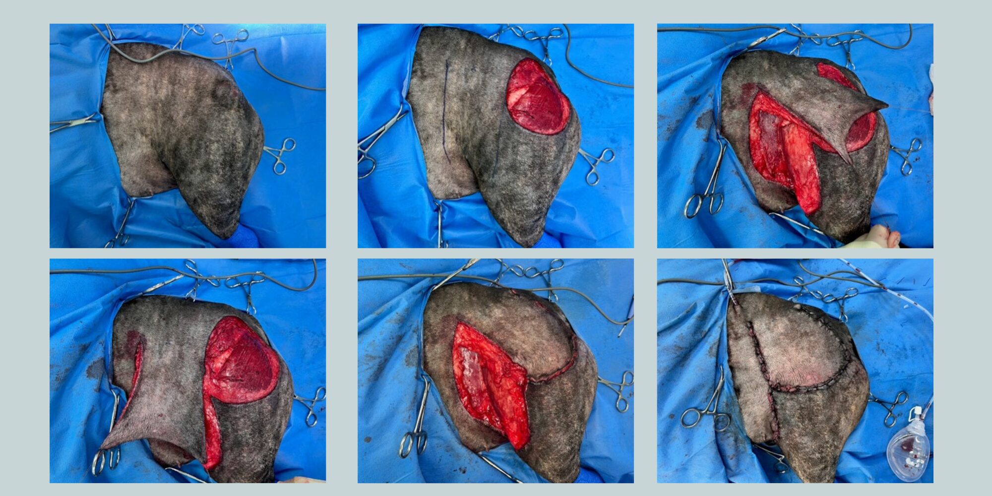
Case example 1: Oncological Reconstruction
Bonnie presented with a three-week history of a 2cm diameter cutaneous mass lesion in her left femoral region. Fine needle aspiration was consistent with a soft tissue sarcoma, thus prompting referral for surgical management. Excision of the mass lesion was performed with 3cm lateral margins and a deep fascial layer. A ventral deep circumflex iliac axial pattern flap was raised and then moved to fill the defect, and an active suction drain was placed in the wound bed before closure.
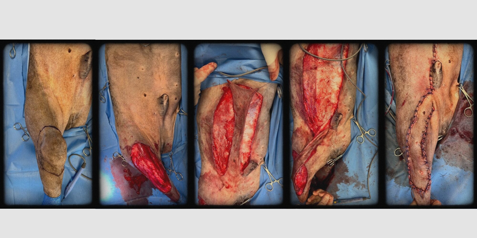
Case example 2. Oncological Reconstruction
Alfie presented with a history of a 3cm diameter subcutaneous mass lesion in his right cranial stifle region. Fine needle aspiration was consistent with a soft tissue sarcoma, thus prompting referral for surgical management. Excision of the mass lesion was performed with 3cm lateral margins and a deep fascial layer. A caudal superficial epigastric axial pattern flap was raised and then moved to fill the defect, and an active suction drain was placed in the wound bed prior to closure.
